Archive 20/12/27

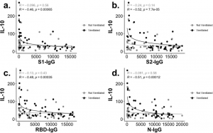
IL-10 and IgG are inversely correlated in the new coronavirus (COVID-19)

A group of Univ. of Virginia Health System has reported interesting phenomena on the expression of IgG, IgA, and IgM in the new coronavirus (COVID-19).
https://www.medrxiv.org/content/10.1101/2020.12.05.20244541v1

IgG, IgM, and IgA are highly expressed as COVID-19 becomes severe.
IL-10 and IgG inversely correlate.


Cytokine IL-10 is usually thought to be an anti-inflammatory one.
IL-10 and IgG are inversely correlated, and moreover, IgG is highly expressed as the disease becomes severe,
Does this mean that IL-10 works as an inflammatory cytokine?

R-type lectin "Sevil" extracted from mussel Mytilisepta virgata strongly binds to asialo-GM1

Lectins with ricin B chains are collectively referred to as R-type lectins.
Ricin is galactose-specific, but the ricin-like structure has an extremely wide biological distribution and has various specificities such as sialic acid, mannose, xylose, etc. in addition to galactose.
The R-type lectin "Sevil" from mussel Mytilisepta virgata binds particularly strongly to the asialo-GM1 structure.
From the Yokohama City University Group,
https://www.nature.com/articles/s41598-020-78926-7

Small molecule compounds that inhibit COVID-19 infection (Protoporphyrin IX (PpIX), Verteporfin): Inhibiting the binding of SARS-CoV-2 to ACE2

A group of Fudan Univ. has shown in vitro that the FDA-approved drugs (Protoporphyrin IX (PpIX), Verteporfin) effectively inhibit the infection of SARS-CoV-2.
This is because these compounds bind to ACE2, inhibiting the binding of SARS-CoV-2 to ACE2.

Clinical trials in vivo are expected.

https://www.sciencedirect.com/science/article/pii/S2095927320307283?via%3Dihub

In chronic HBV, changes are occurring in expression of C-type lectin receptors on dendritic cells

Dendritic cells (DCs) are at the forefront of the immune reaction.
When pathogens such as viruses invade, Toll-like receptors (TLR) and C-type lectin receptors (CLRs) expressed on DCs recognize the molecular structure peculiar to pathogens and trigger immune responses.
The CLR of DCs includes DCIR/CD367/CLEC4A, DECTIN1/CD369/CLEC7A, DECTIN2/CLEC6A, DNGR1/CD370/CLEC9A, MMR/CD206, DEC205/CD205, DC-SIGN/CD209, langerin/CD207, BDCA2/CD303/CLEC4C, etc.
The function of these lectins remain largely unknow, but for example,
DCIR binds to high mannose/fucose glycans and works inhibitory on the secretion of IL12 and TNFα, while DECTIN1 recognizes β-1,3-glucans and conversely promotes the secretion of these inflammatory cytokines.

A group of Univ. Grenoble Alpes discussed how the expression of various CLRs changes in chronic HBV based on experimental results performed using Flow cytometry.
https://onlinelibrary.wiley.com/doi/10.1002/cti2.1208

There are three subclasses of DC: CD1c/BDCA1 (cDC2s), CD141/BDCA3 (cDC1s), and plasmacytoid DCs (pDCs).

    Blood cDC2s: DECTIN1, MMR decreased,
    Liver cDC2s: DCIR, MMR decreased,
    Blood cDC1s: DECTIN1, CLEC9A decreased, Fcɣ Receptor increased slightly,
    Liver cDC1s: DCIR, CLEC9A, Fcɣ receptor, MMR decreased, DECTIN1 increased slightly,
    Blood pCDs: Little change,
    Liver pCDs: DCIR increased, Fcε receptor increased slightly.

Such a change is happening.

Although the changes are very complex, it is worth noting that chronic HBV causes changes in the expression of CLRs in DCs.
Overall, CLR expression seems to be decreasing, which makes it easier for HBV to escape from immune system, right?
A new exosome marker for Alzheimer’s disease was discovered

A group of AIST and Kyoto Univ. has discovered a high-performance novel exosome marker as a novel marker for Alzheimer's disease.
https://pubmed.ncbi.nlm.nih.gov/33345458/

They found that in patients with Alzheimer's disease, there was a significant difference in glycan modification of exosomes compared to healthy people, and the exosomes were highly high mannosylated.
Using a high mannose specific lectin, its lectin blotting revealed a very strong band around 80 kDa, which was identified as a marker of exosomes, CD61.

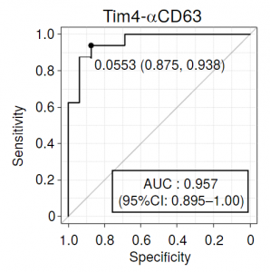
A sandwich assay (Tim4-αCD63) assembled with an antibody to the exosome marker CD63 and Tim4 (T cell immunoglobulin and mucin domain-containing protein 4) showed the highest discriminating ability among (CD61, CD41, CD63, and CD9), and the obtained AUC reached 0.957.

Effects of statins on the new coronavirus (COVID-19)

Statins are widely used as a drug to lower cholesterol.
In the case of viral infection, it is well inferred that changes in cholesterol and sphingolipids may affect the infection of the virus, since lipid rafts present on the cell membrane are intermingling in the process of infection.
https://www.sciencedirect.com/science/article/pii/S0306987720333430?via%3Dihub

In COVID-19, it was found that the severe Hazard Ratio (HR) decreased significantly with statins,
HR=0.70 (95% CI: 0.53 – 0.94).

Similarly, the effect of statins has been also recognized in influenza,
Odds ratio (OR)=0.72 (95% CI: 0.38 – 1.33).

But, further research is needed to clarify if statins inhibit viral infection itself or inhibit the replication of the virus.

RNA mutations in the new coronavirus (SARS-CoV-2) mutant strain (B.1.1.7), which is all the rage in the UK

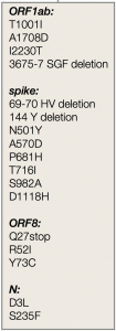
There is a recent report on RNA mutations in the new coronavirus (SARS-CoV-2) mutant strain (B.1.1.7), which is all the rage in the UK.

https://virological.org/t/preliminary-genomic-characterisation-of-an-emergent-sars-cov-2-lineage-in-the-uk-defined-by-a-novel-set-of-spike-mutations/563

There are 14 non-synonymous mutations, 6 synonymous mutations and 3 deletions, and it seems to be a fairly large mutation.

Fc-fusion ivermectin-containing nanoparticle drug that can be administered orally for the new coronavirus (COVID-19)

Ivermectin, an antiparasitic drug, has been known to have antiviral effects, too. The mechanism of action is thought to be because inhibiting nuclear transport proteins such as an importin prevents the transport of viral proteins into the nucleus.
Ivermectin itself is toxic, and EC50 is 1 to 10 mM. When ivermectin is administered to the human body with 150 µg/kg of dose, the concentration in plasma is only 9 to 75 ng/mL, and the concentration is too low to be effective.
Therefore, the following group has verified the effect of a therapeutic agent (Fc-fusion ivermectin-containing nanoparticles that can be administered orally: T-Fc-IVM-NPs) that enables intestinal absorption of T-Fc-IVM-NPs via interaction between FcRn receptors expressed on gut epithelial cells and Fc on ivermectin-containing nanoparticles.
https://pubs.acs.org/doi/10.1021/acsptsci.0c00179

At this stage, although it was an in vitro experiment using HEK293 infected with the new novel coronavirus (SARS-CoV-2), it is said that the expression of ACE2 and the expression of S-protein were suppressed by administering T-Fc-IVM-NPs. In addition, this method can be expected to be effective against other viruses.

It’s good, isn’t it?

Macrophages are strongly involved in severity of the new coronavirus (COVID-19)

In the new coronavirus (COVID-19), there is a large amount of macrophages in the bronchoalveolar lavage fluid, suggesting that macrophages are greatly involved in the severity. Macrophages are not just phagocytes, they have many subsets, and each has its own functions.

The following group has reported that transcription factors (MAFB and MAF) in macrophages are significantly related to the progression of COVID-19.

https://www.frontiersin.org/articles/10.3389/fimmu.2020.603507/full

MAFB, a transcription factor, promotes a subset of pathogenic pro-fibrotic SPP1+ macrophages, while transcription factor MAF has the function of suppressing inflammatory FCN1+ macrophage subsets. When COVID-19 becomes more severe, the MAFB/MAF ratio increases. In other words, subsets of macrophages are controlled in a direction that accelerates inflammation of lung tissue and simultaneously accelerates fibrosis.
Therefore, they have concluded that it might be effective to silence MAFB and induce overexpression of MAF when treating COVID-19.

Efficacy of MP (methyl-prednisolone 1000mg for three days) pulse therapy in severe periods from a COVID-19 case report

The blog management is not a medical doctor, but the following case report may be useful if you infected with the novel coronavirus (COVID-19) and become severe.
https://www.sciencedirect.com/science/article/pii/S2213007120305323?via%3Dihub

In general, steroids are commonly used to reduce inflammation, but long-term use has significant side effects and slows the elimination of the virus in COVID-19.
However, in the late phase of covid-19, there were four cases in which MP (methyl-prednisolone) was highly effective when its pulse therapy was performed at high concentrations for a short period of time (1000 mg, 3 days).
All have recovered and have been discharged from hospital.

It may be good to tell your family.

Powered by WordPress |Copyright © 2020 Emukk. All rights reserved